Copyright
©2012 Baishideng Publishing Group Co.
World J Gastroenterol. Nov 7, 2012; 18(41): 5994-5998
Published online Nov 7, 2012. doi: 10.3748/wjg.v18.i41.5994
Published online Nov 7, 2012. doi: 10.3748/wjg.v18.i41.5994
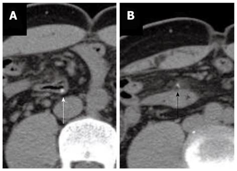
Figure 2 Two follow-up unenhanced abdominal computer tomography images, which reveal the radiopaque shadow still lodged in the intestinal segment.
The fish bone rotates and becomes parallel to the distal ileum lumen. A: Most of the fish bone is still inside the intestinal lumen (white arrow). One end of the fish bone penetrates out the intestinal wall into the mesenteric fat; B: Minimal local inflammatory infiltration contains the protruding part. No free air or abscess is noted (black arrow). The distance between these two images is 18 mm.
- Citation: Kuo CC, Jen TK, Wen CH, Liu CP, Hsiao HS, Liu YC, Chen KH. Medical treatment for a fish bone-induced ileal micro-perforation: A case report. World J Gastroenterol 2012; 18(41): 5994-5998
- URL: https://www.wjgnet.com/1007-9327/full/v18/i41/5994.htm
- DOI: https://dx.doi.org/10.3748/wjg.v18.i41.5994


