Copyright
©2012 Baishideng Publishing Group Co.
World J Gastroenterol. Nov 7, 2012; 18(41): 5990-5993
Published online Nov 7, 2012. doi: 10.3748/wjg.v18.i41.5990
Published online Nov 7, 2012. doi: 10.3748/wjg.v18.i41.5990
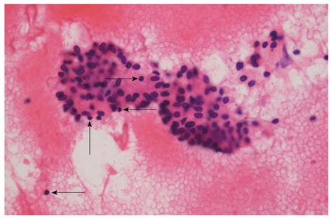
Figure 3 Photomicrograph of cytologic specimen obtained by endoscopic ultrasound-fine needle aspiration, showing lymphocytes (arrows) and irregular sheets of bland ductal epithelial cells on the bloody background (hematoxylin and eosin, × 400).
- Citation: Kim JH, Chang JH, Nam SM, Lee MJ, Maeng IH, Park JY, Im YS, Kim TH, Kim CW, Han SW. Newly developed autoimmune cholangitis without relapse of autoimmune pancreatitis after discontinuing prednisolone. World J Gastroenterol 2012; 18(41): 5990-5993
- URL: https://www.wjgnet.com/1007-9327/full/v18/i41/5990.htm
- DOI: https://dx.doi.org/10.3748/wjg.v18.i41.5990


