Copyright
©2012 Baishideng Publishing Group Co.
World J Gastroenterol. Oct 28, 2012; 18(40): 5830-5832
Published online Oct 28, 2012. doi: 10.3748/wjg.v18.i40.5830
Published online Oct 28, 2012. doi: 10.3748/wjg.v18.i40.5830
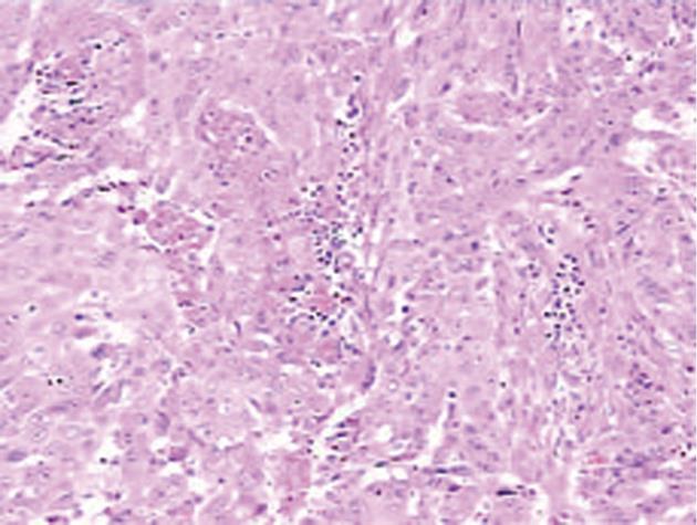
Figure 3 Histology of the tumor: Squamous cells with keratinization and areas of necrosis.
Hematoxylin-eosin stain, × 10.
- Citation: Zhu KL, Li DY, Jiang CB. Primary squamous cell carcinoma of the liver associated with hepatolithiasis: A case report. World J Gastroenterol 2012; 18(40): 5830-5832
- URL: https://www.wjgnet.com/1007-9327/full/v18/i40/5830.htm
- DOI: https://dx.doi.org/10.3748/wjg.v18.i40.5830


