Copyright
©2012 Baishideng Publishing Group Co.
World J Gastroenterol. Oct 28, 2012; 18(40): 5816-5820
Published online Oct 28, 2012. doi: 10.3748/wjg.v18.i40.5816
Published online Oct 28, 2012. doi: 10.3748/wjg.v18.i40.5816
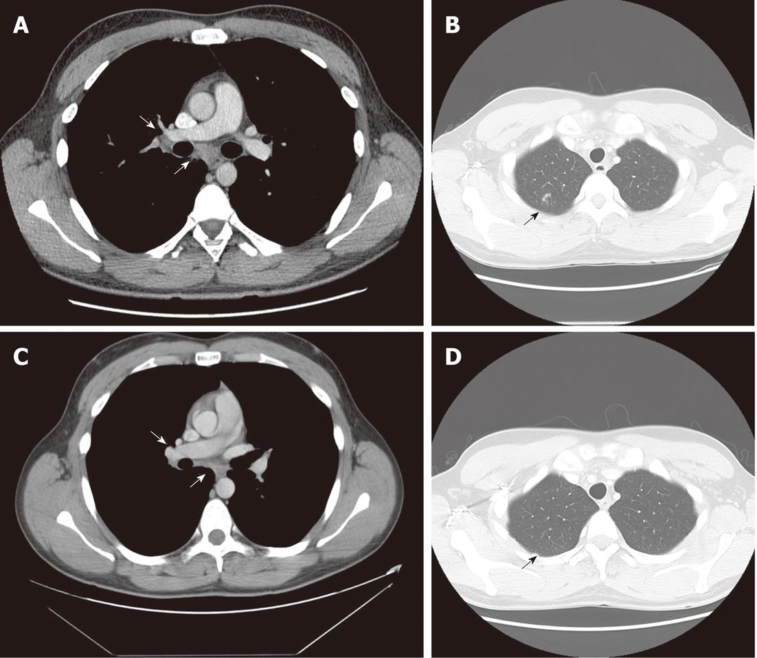
Figure 4 Thoracic computer tomographic scans of mediastinal, hilar lymph nodes and pulmonary nodules.
A significant decrease in size of mediastinal and hilar lymph nodes (A, white arrows) is shown before initiation of antiviral treatment but pulmonary nodules remain at the same levels (B, black arrow; October 2009). At the end of treatment no progression of lymph nodes is revealed (C, white arrows) and pulmonary nodules are absorbed (D, black arrow; December 2010).
- Citation: Brjalin V, Salupere R, Tefanova V, Prikk K, Lapidus N, Jõeste E. Sarcoidosis and chronic hepatitis C: A case report. World J Gastroenterol 2012; 18(40): 5816-5820
- URL: https://www.wjgnet.com/1007-9327/full/v18/i40/5816.htm
- DOI: https://dx.doi.org/10.3748/wjg.v18.i40.5816


