Copyright
©2012 Baishideng Publishing Group Co.
World J Gastroenterol. Oct 28, 2012; 18(40): 5816-5820
Published online Oct 28, 2012. doi: 10.3748/wjg.v18.i40.5816
Published online Oct 28, 2012. doi: 10.3748/wjg.v18.i40.5816
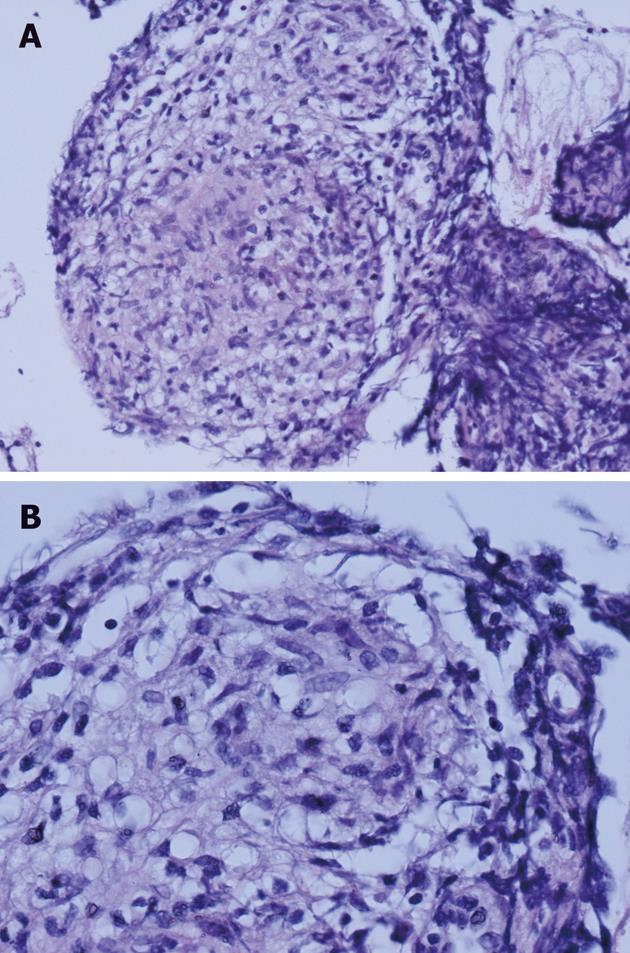
Figure 2 Biopsy taken from a pulmonary lymph node.
Two tight naked round well-formed granulomas are surrounded by a small amount of lymphocytic infiltration. The granulomas consist of epithelioid cells; there are no necrosis, giant cells, Shaumann or asteroid bodies. Stained with hematoxylin and eosin, magnification × 200 (A) and × 400 (B).
- Citation: Brjalin V, Salupere R, Tefanova V, Prikk K, Lapidus N, Jõeste E. Sarcoidosis and chronic hepatitis C: A case report. World J Gastroenterol 2012; 18(40): 5816-5820
- URL: https://www.wjgnet.com/1007-9327/full/v18/i40/5816.htm
- DOI: https://dx.doi.org/10.3748/wjg.v18.i40.5816


