Copyright
©2012 Baishideng Publishing Group Co.
World J Gastroenterol. Oct 28, 2012; 18(40): 5695-5701
Published online Oct 28, 2012. doi: 10.3748/wjg.v18.i40.5695
Published online Oct 28, 2012. doi: 10.3748/wjg.v18.i40.5695
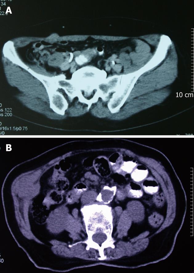
Figure 2 Computerized tomography scans of subcutaneous mass.
A: A 2 cm × 1.5 cm subcutaneous mass at right lateral abdominal wall, which near a trocar scar on physical examination; B: A 4 cm × 3 cm × 3 cm subcutaneous mass at right lateral abdominal wall.
- Citation: Liu QD, Chen JZ, Xu XY, Zhang T, Zhou NX. Incidence of port-site metastasis after undergoing robotic surgery for biliary malignancies. World J Gastroenterol 2012; 18(40): 5695-5701
- URL: https://www.wjgnet.com/1007-9327/full/v18/i40/5695.htm
- DOI: https://dx.doi.org/10.3748/wjg.v18.i40.5695


