Copyright
©2012 Baishideng Publishing Group Co.
World J Gastroenterol. Jan 28, 2012; 18(4): 356-367
Published online Jan 28, 2012. doi: 10.3748/wjg.v18.i4.356
Published online Jan 28, 2012. doi: 10.3748/wjg.v18.i4.356
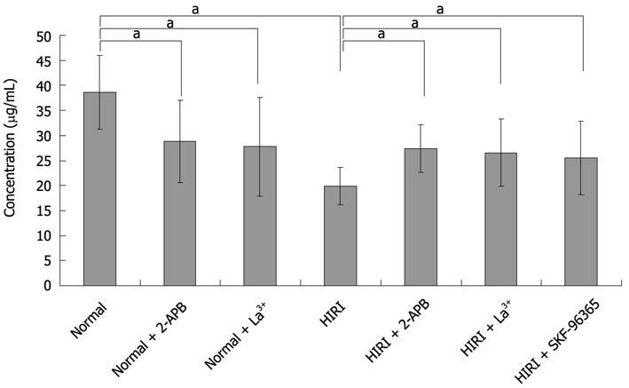
Figure 7 Taurocholate measurements in hepatic ischemia-reperfusion injuried hepatocytes.
Bar graph of taurocholate secreted by hepatocytes is distinctly higher in the supernatant of cultured normal hepatocytes (38.58 ± 7.35 μg/mL, n = 6) than for cells after exposure to 100 μmol/L 2-APB (28.85 ± 8.18 μg/mL, aP < 0.05, n = 6), 100 μmol/L La3+ (27.76 ± 9.86 μg/mL, aP < 0.05, n = 6) or HIRI hepatocytes (19.92 ± 3.75 μg/mL, aP < 0.05, n = 6). Whereas 100 μmol/L 2-APB, 100 μmol/L La3+ and 10 μmol/L SKF-96365 could respectively reverse the taurocholate level to 27.42 ± 4.74, 26.58 ± 6.67 and 25.52 ± 7.30 μg/mL in HIRI hepatocytes, aP < 0.05, n = 6. HIRI: Hepatic ischemia-reperfusion injury; 2-APB: 2-aminoethoxydiphenyl borate.
- Citation: Pan LJ, Zhang ZC, Zhang ZY, Wang WJ, Xu Y, Zhang ZM. Effects and mechanisms of store-operated calcium channel blockade on hepatic ischemia-reperfusion injury in rats. World J Gastroenterol 2012; 18(4): 356-367
- URL: https://www.wjgnet.com/1007-9327/full/v18/i4/356.htm
- DOI: https://dx.doi.org/10.3748/wjg.v18.i4.356


