Copyright
©2012 Baishideng Publishing Group Co.
World J Gastroenterol. Oct 21, 2012; 18(39): 5645-5648
Published online Oct 21, 2012. doi: 10.3748/wjg.v18.i39.5645
Published online Oct 21, 2012. doi: 10.3748/wjg.v18.i39.5645
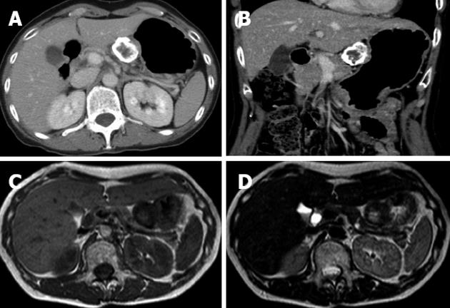
Figure 2 Computed tomography and magnetic resonance imaging findings.
Contrast-enhanced axial (A) and coronal (B) computed tomography examination demonstrated that the marginal zone of the tumor was calcified, and that the internal portion of the tumor was enhanced heterogeneously. Magnetic resonance imaging T1-weighted image (C) and T2-weighted image (D) revealed a low intensity marginal zone of tumor reflecting calcification.
- Citation: Izawa N, Sawada T, Abiko R, Kumon D, Hirakawa M, Kobayashi M, Obinata N, Nomoto M, Maehata T, Yamauchi SI, Kouro T, Tsuda T, Kitajima S, Yasuda H, Tanaka K, Tanaka I, Hoshikawa M, Takagi M, Itoh F. Gastrointestinal stromal tumor presenting with prominent calcification. World J Gastroenterol 2012; 18(39): 5645-5648
- URL: https://www.wjgnet.com/1007-9327/full/v18/i39/5645.htm
- DOI: https://dx.doi.org/10.3748/wjg.v18.i39.5645


