Copyright
©2012 Baishideng Publishing Group Co.
World J Gastroenterol. Oct 14, 2012; 18(38): 5479-5484
Published online Oct 14, 2012. doi: 10.3748/wjg.v18.i38.5479
Published online Oct 14, 2012. doi: 10.3748/wjg.v18.i38.5479
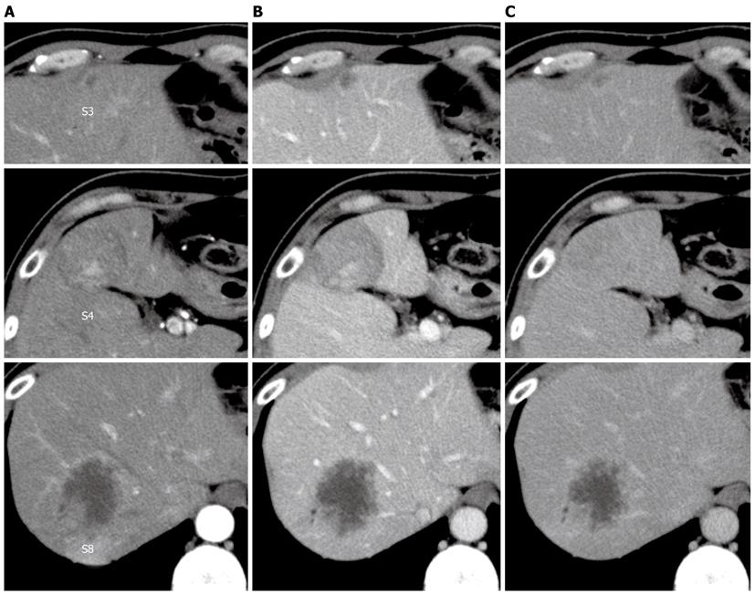
Figure 1 Dynamic computed tomography of the liver.
A: Arterial phase; B: Portal phase; C: Venous phase. A hypo- or iso-dense tumor with heterogeneous enhancement in segment 4, a hypodense tumor with peripheral enhancement in segment 8, and a small hypodense tumor in segment 3.
- Citation: Takehara K, Aoki H, Takehara Y, Yamasaki R, Tanakaya K, Takeuchi H. Primary hepatic leiomyosarcoma with liver metastasis of rectal cancer. World J Gastroenterol 2012; 18(38): 5479-5484
- URL: https://www.wjgnet.com/1007-9327/full/v18/i38/5479.htm
- DOI: https://dx.doi.org/10.3748/wjg.v18.i38.5479


