Copyright
©2012 Baishideng Publishing Group Co.
World J Gastroenterol. Oct 14, 2012; 18(38): 5476-5478
Published online Oct 14, 2012. doi: 10.3748/wjg.v18.i38.5476
Published online Oct 14, 2012. doi: 10.3748/wjg.v18.i38.5476
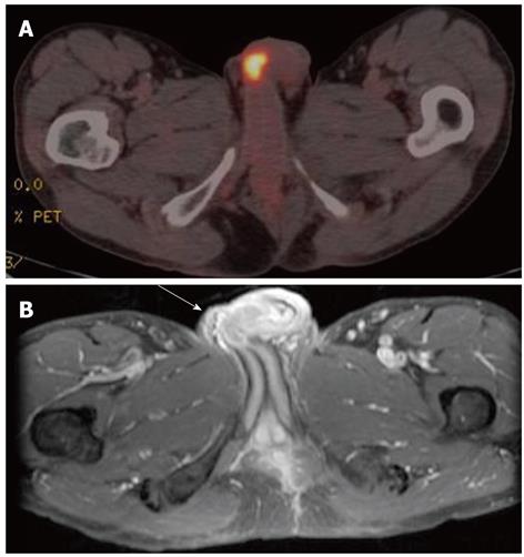
Figure 1 Positron emission tomography and magnetic resonance imaging image.
A: Positron emission tomography/computed tomography showing high 18-F fluorodeoxyglucose uptake in the penis; B: Gadolinium-enhanced fat-suppressed T1-weighted image showing a low intensity lesion on the middle penis shaft (white arrow).
- Citation: Kimura Y, Shida D, Nasu K, Matsunaga H, Warabi M, Inoue S. Metachronous penile metastasis from rectal cancer after total pelvic exenteration. World J Gastroenterol 2012; 18(38): 5476-5478
- URL: https://www.wjgnet.com/1007-9327/full/v18/i38/5476.htm
- DOI: https://dx.doi.org/10.3748/wjg.v18.i38.5476


