Copyright
©2012 Baishideng Publishing Group Co.
World J Gastroenterol. Oct 14, 2012; 18(38): 5360-5368
Published online Oct 14, 2012. doi: 10.3748/wjg.v18.i38.5360
Published online Oct 14, 2012. doi: 10.3748/wjg.v18.i38.5360
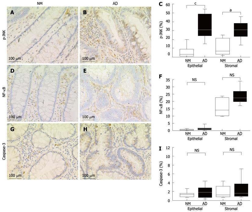
Figure 3 Immunohistochemical analyses in the normal colorectal mucosa and adenoma tissues.
A: Phospho-c-Jun N-terminal kinase (p-JNK) expression in the normal colorectal mucosa; B: p-JNK expression in the adenoma tissues; C: The percentage of p-JNK positive cells; D: Nuclear factor-κ B (NF-κB) expression in the normal colorectal mucosa; E: NF-κB expression in the adenoma tissues; F: The percentage of NF-κB-positive cells; G: Caspase-3 expression in the normal colorectal mucosa; H: Caspase-3 expression in the adenoma tissues; I: The percentage of caspase-3-positive cells. Box plots display median values and interquartile ranges (C, F, I). The non-outlier range is also shown. aP < 0.05 between NM and AD in stromal of p-JNK; cP < 0.05 between NM and AD in epithelial of p-JNK. NS: Non-significant; NM: Normal mucosa; AD: Adenoma.
- Citation: Hosono K, Yamada E, Endo H, Takahashi H, Inamori M, Hippo Y, Nakagama H, Nakajima A. Increased tumor necrosis factor receptor 1 expression in human colorectal adenomas. World J Gastroenterol 2012; 18(38): 5360-5368
- URL: https://www.wjgnet.com/1007-9327/full/v18/i38/5360.htm
- DOI: https://dx.doi.org/10.3748/wjg.v18.i38.5360


