Copyright
©2012 Baishideng Publishing Group Co.
World J Gastroenterol. Oct 7, 2012; 18(37): 5211-5218
Published online Oct 7, 2012. doi: 10.3748/wjg.v18.i37.5211
Published online Oct 7, 2012. doi: 10.3748/wjg.v18.i37.5211
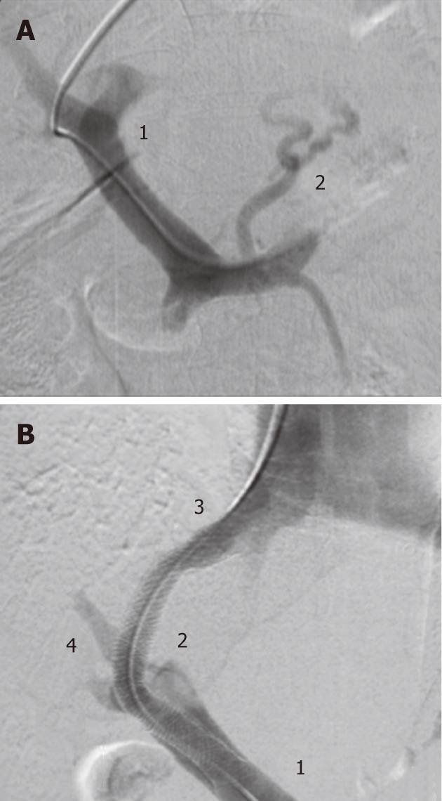
Figure 1 Fluoroscopic images showing transjugular intrahepatic portosystemic shunt placement procedure.
A: Portogram after catheterisation of the portal vein, showing perfusion of the portal vein system (1) and oesophageal varices (2); B: Portogram after transjugular intrahepatic portosystemic stent placement. Contrast can be seen in the portal vein (1), through the shunt (2) flowing into the hepatic vein and inferior vena cava (3). Decompression of the portosystemic pressure can be seen in reduced contrast in the portal branch (4). The varices can no longer be identified in the fluoroscopic image.
- Citation: Heinzow HS, Lenz P, Köhler M, Reinecke F, Ullerich H, Domschke W, Domagk D, Meister T. Clinical outcome and predictors of survival after TIPS insertion in patients with liver cirrhosis. World J Gastroenterol 2012; 18(37): 5211-5218
- URL: https://www.wjgnet.com/1007-9327/full/v18/i37/5211.htm
- DOI: https://dx.doi.org/10.3748/wjg.v18.i37.5211


