Copyright
©2012 Baishideng Publishing Group Co.
World J Gastroenterol. Oct 7, 2012; 18(37): 5181-5187
Published online Oct 7, 2012. doi: 10.3748/wjg.v18.i37.5181
Published online Oct 7, 2012. doi: 10.3748/wjg.v18.i37.5181
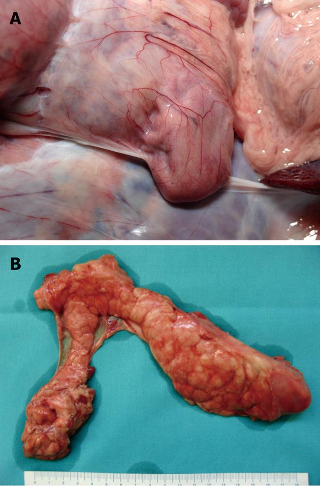
Figure 2 Gross anatomy of the porcine pancreas after double-balloon enteroscopy.
A: In situ image of the left lobe (tail); B: Aspect of the whole pancreas immediately after removal from cadaver.
- Citation: Latorre R, Soria F, López-Albors O, Sarriá R, Sánchez-Margallo F, Esteban P, Carballo F, Pérez-Cuadrado E. Effect of double-balloon enteroscopy on pancreas: An experimental porcine model. World J Gastroenterol 2012; 18(37): 5181-5187
- URL: https://www.wjgnet.com/1007-9327/full/v18/i37/5181.htm
- DOI: https://dx.doi.org/10.3748/wjg.v18.i37.5181


