Copyright
©2012 Baishideng Publishing Group Co.
World J Gastroenterol. Sep 28, 2012; 18(36): 5042-5050
Published online Sep 28, 2012. doi: 10.3748/wjg.v18.i36.5042
Published online Sep 28, 2012. doi: 10.3748/wjg.v18.i36.5042
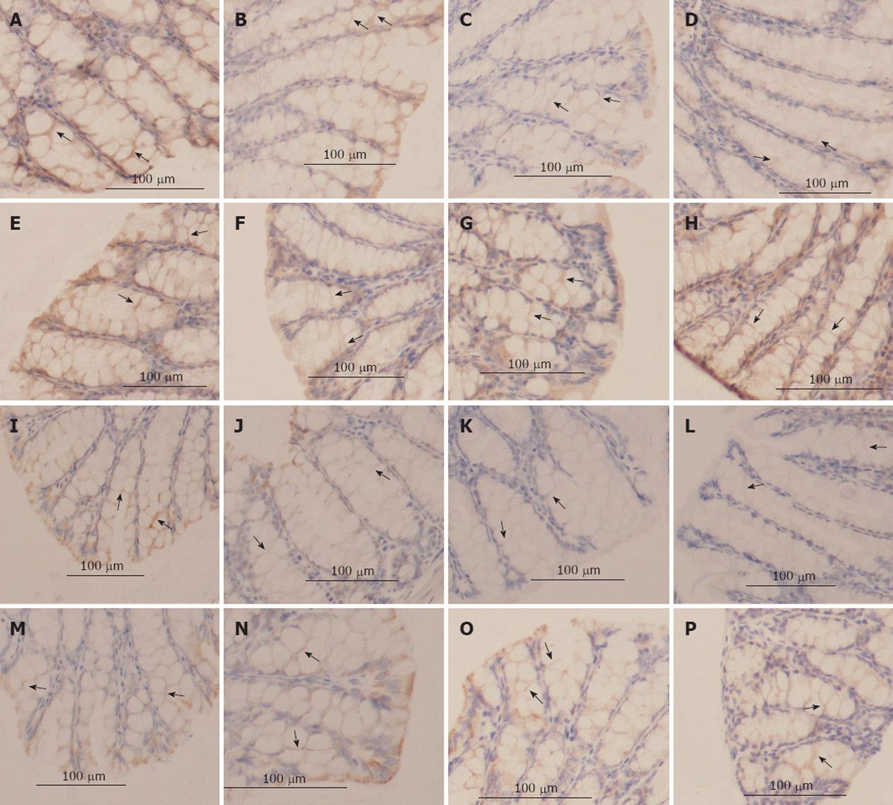
Figure 3 The tight junction proteins, occludin and claudin-1 determined by immunohistochemistry (400×).
A: Occludin staining in the saline group; B-D: Occludin staining 2 h, 6 h, and 9 h after injection in the D-galactosamine (GalN)/lipopolysaccharide (LPS)-treated mice; E-H: Occludin staining 6 h after injection in the anti-tumor necrosis factor alpha (TNF-α) immunoglobulin G (IgG) antibody pretreated group within the TNF-α-treated group; the LPS-treated group and the D-GalN-treated group, respectively; I: Claudin-1 staining in the saline group; J-L: Claudin-1 staining 2, 6, and 9 h after injection in the GalN/LPS-treated mice; M-P: Claudin-1 staining 6 h after injection in the anti-TNF-α IgG antibody pretreated group, the TNF-α-treated group, the LPS-treated group and the D-GalN-treated group, respectively. The mucosal tissue sections were double-labeled for proteins (brown color). Labeled sections were analyzed immunohisto-chemically. Decreased protein staining in the epithelial cells were observed at 2-9 h after injection in the GalN/LPS-treated mice and the TNF-α-treated group, and was absent in the other controls and the antibody pretreated group.
- Citation: Li GZ, Wang ZH, Cui W, Fu JL, Wang YR, Liu P. Tumor necrosis factor alpha increases intestinal permeability in mice with fulminant hepatic failure. World J Gastroenterol 2012; 18(36): 5042-5050
- URL: https://www.wjgnet.com/1007-9327/full/v18/i36/5042.htm
- DOI: https://dx.doi.org/10.3748/wjg.v18.i36.5042


