Copyright
©2012 Baishideng Publishing Group Co.
World J Gastroenterol. Sep 21, 2012; 18(35): 4967-4972
Published online Sep 21, 2012. doi: 10.3748/wjg.v18.i35.4967
Published online Sep 21, 2012. doi: 10.3748/wjg.v18.i35.4967
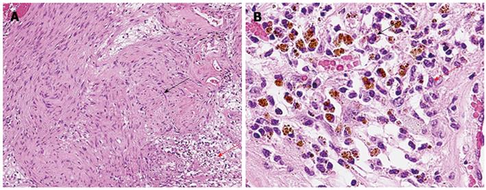
Figure 4 Microscopic examination.
A: B-mode ultrasound. Microscopic examination revealed that Antoni A hypercellular area (black arrow) and Antoni B hypocellular area (red arrow) existed together in a complex; B: Vascular phase. In the Antoni B region, there were edematous stroma, vasodilatation, aggregation of siderophores (black arrow), and slight permeation of chronic inflammatory cells of the histiocyte and lymphocyte types (red arrow).
- Citation: Ota Y, Aso K, Watanabe K, Einama T, Imai K, Karasaki H, Sudo R, Tamaki Y, Okada M, Tokusashi Y, Kono T, Miyokawa N, Haneda M, Taniguchi M, Furukawa H. Hepatic schwannoma: Imaging findings on CT, MRI and contrast-enhanced ultrasonography. World J Gastroenterol 2012; 18(35): 4967-4972
- URL: https://www.wjgnet.com/1007-9327/full/v18/i35/4967.htm
- DOI: https://dx.doi.org/10.3748/wjg.v18.i35.4967


