Copyright
©2012 Baishideng Publishing Group Co.
World J Gastroenterol. Sep 21, 2012; 18(35): 4967-4972
Published online Sep 21, 2012. doi: 10.3748/wjg.v18.i35.4967
Published online Sep 21, 2012. doi: 10.3748/wjg.v18.i35.4967
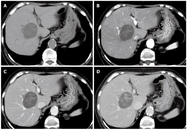
Figure 1 Abdominal computed tomography.
A: Plain; B: Arterial dominant phase; C: Portal venous phase; D: Late venous phase. Abdominal computed tomography (CT) showed an approximately 5-cm well-defined round tumor in segment 8 of the right hepatic lobe. CT showed heterogeneous enhancement in the arterial dominant phase; and delayed the enhancement until the portal venous and late venous phases.
- Citation: Ota Y, Aso K, Watanabe K, Einama T, Imai K, Karasaki H, Sudo R, Tamaki Y, Okada M, Tokusashi Y, Kono T, Miyokawa N, Haneda M, Taniguchi M, Furukawa H. Hepatic schwannoma: Imaging findings on CT, MRI and contrast-enhanced ultrasonography. World J Gastroenterol 2012; 18(35): 4967-4972
- URL: https://www.wjgnet.com/1007-9327/full/v18/i35/4967.htm
- DOI: https://dx.doi.org/10.3748/wjg.v18.i35.4967


