Copyright
©2012 Baishideng Publishing Group Co.
World J Gastroenterol. Sep 21, 2012; 18(35): 4925-4933
Published online Sep 21, 2012. doi: 10.3748/wjg.v18.i35.4925
Published online Sep 21, 2012. doi: 10.3748/wjg.v18.i35.4925
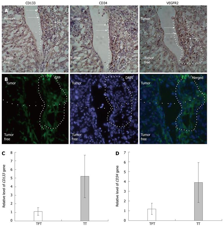
Figure 2 Recruitment of endothelial progenitor cells to hepatocellular carcinoma tissue.
A: Distribution endothelial progenitor cells (EPCs) in tumor and tumor-free tissue in four consecutive 2 μm sections (arrows: Cells expressing the indicated antigens and recruited to tumors and tumor vessels); B: Bone-marrow cells labeled by green fluorescent protein recruited to tumors and tumor vessels, occupying the same position as antigen positive cells [blue regions: Nuclei stained by DAPI (frozen section, 2 μm, magnification 200)]; C: Relative levels of CD133 mRNA in tumor tissue (n = 7) and tumor-free tissue (n = 6); D: Relative level of CD34 mRNA in tumor tissue (n = 7) and tumor-free tissue (n = 7). TFT: Tumor-free tissues; TT: Tumor tissues; DAPI: 4',6'-diamidino-2-phenylindole hydrochloride; GFP: Green fluorescent protein.
- Citation: Sun XT, Yuan XW, Zhu HT, Deng ZM, Yu DC, Zhou X, Ding YT. Endothelial precursor cells promote angiogenesis in hepatocellular carcinoma. World J Gastroenterol 2012; 18(35): 4925-4933
- URL: https://www.wjgnet.com/1007-9327/full/v18/i35/4925.htm
- DOI: https://dx.doi.org/10.3748/wjg.v18.i35.4925


