Copyright
©2012 Baishideng Publishing Group Co.
World J Gastroenterol. Sep 21, 2012; 18(35): 4905-4911
Published online Sep 21, 2012. doi: 10.3748/wjg.v18.i35.4905
Published online Sep 21, 2012. doi: 10.3748/wjg.v18.i35.4905
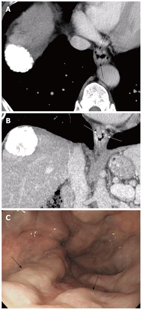
Figure 1 Imaging findings are shown for grade 2 esophageal varices in the distal esophagus in a 61-year-old man with hepatocellular carcinoma and cirrhosis.
A: A transversecomputed tomography scan shows distinct enhancing nodular lesions (arrows) protruding into the luminal space. All observers recorded the lesions as high-risk esophageal varices. Periesophageal varices are not found; B: On coronal images, distinct enhancing nodular lesions (arrows) are noted, and all observers considered the lesions as high-risk esophageal varices; C: A conventional endoscopic image confirms the presence of beaded esophageal varices (arrows).
- Citation: Kim H, Choi D, Lee JH, Lee SJ, Jo H, Gwak GY, Koh KC, Choi MS, Kim S. High-risk esophageal varices in patients treated with locoregional therapy for hepatocellular carcinoma: Assessment with liver computed tomography. World J Gastroenterol 2012; 18(35): 4905-4911
- URL: https://www.wjgnet.com/1007-9327/full/v18/i35/4905.htm
- DOI: https://dx.doi.org/10.3748/wjg.v18.i35.4905


