Copyright
©2012 Baishideng Publishing Group Co.
World J Gastroenterol. Sep 7, 2012; 18(33): 4549-4556
Published online Sep 7, 2012. doi: 10.3748/wjg.v18.i33.4549
Published online Sep 7, 2012. doi: 10.3748/wjg.v18.i33.4549
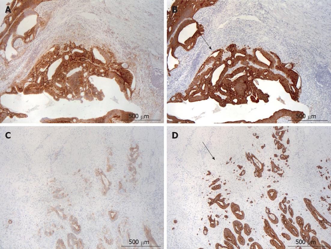
Figure 1 The immunohistochemistry for CD44 variant 6 and Pan-cytokeratin.
A: The same tumor with “front-positive” membranous staining of CD44 variant 6 (CD44v6); B: Expanding growth pattern as shown with Pan-cytokeratin (Ck-Pan) staining; C: The same tumor with “front-negative” membranous staining of CD44v6; D: Infiltrating growth pattern as shown with Ck-Pan staining. Invasive front is indicated with arrows.
- Citation: Avoranta ST, Korkeila EA, Syrjänen KJ, Pyrhönen SO, Sundström JTT. Lack of CD44 variant 6 expression in rectal cancer invasive front associates with early recurrence. World J Gastroenterol 2012; 18(33): 4549-4556
- URL: https://www.wjgnet.com/1007-9327/full/v18/i33/4549.htm
- DOI: https://dx.doi.org/10.3748/wjg.v18.i33.4549


