Copyright
©2012 Baishideng Publishing Group Co.
World J Gastroenterol. Aug 28, 2012; 18(32): 4317-4322
Published online Aug 28, 2012. doi: 10.3748/wjg.v18.i32.4317
Published online Aug 28, 2012. doi: 10.3748/wjg.v18.i32.4317
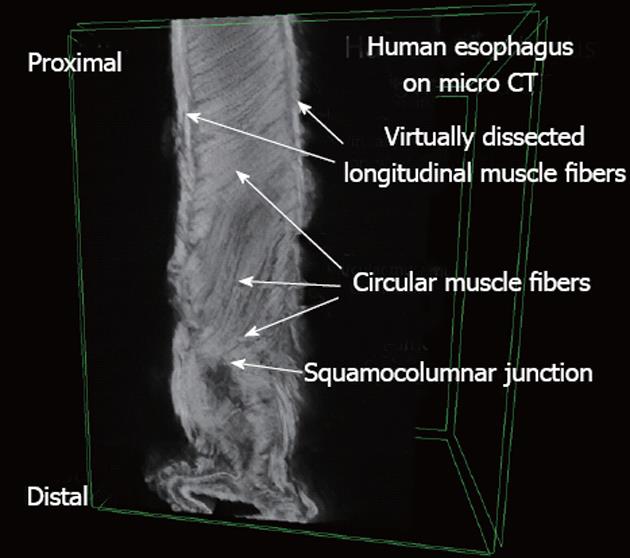
Figure 2 A human esophagus obtained using micro-computed tomography.
The longitudinal muscle fibers were virtually dissected to expose the underlying circular muscle fibers. Measurements were made starting from the squamocolumnar junction.
- Citation: Vegesna AK, Chuang KY, Besetty R, Phillips SJ, Braverman AS, Barbe MF, Ruggieri MR, Miller LS. Circular smooth muscle contributes to esophageal shortening during peristalsis. World J Gastroenterol 2012; 18(32): 4317-4322
- URL: https://www.wjgnet.com/1007-9327/full/v18/i32/4317.htm
- DOI: https://dx.doi.org/10.3748/wjg.v18.i32.4317


