Copyright
©2012 Baishideng Publishing Group Co.
World J Gastroenterol. Aug 21, 2012; 18(31): 4228-4232
Published online Aug 21, 2012. doi: 10.3748/wjg.v18.i31.4228
Published online Aug 21, 2012. doi: 10.3748/wjg.v18.i31.4228
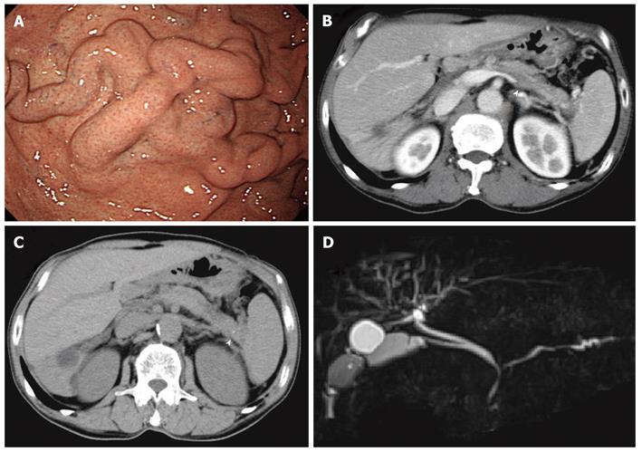
Figure 3 Autoimmune pancreatitis that did not improve with steroid therapy.
A: Esophagogastoroduodenoscopy showed gastric varices in the fundus of the stomach; B: Computed tomography (CT) showed a slightly enlarged pancreas with a capsule-like rim, an obstructed splenic vein (arrowhead), and splenomegaly; C: CT showed a pancreatic stone in the pancreatic tail (arrowhead); D: Magnetic resonance cholangiopancreatography showed irregular narrowing of the main pancreatic duct in the pancreatic head and body, slight dilation of the main pancreatic duct in the pancreatic tail, stricture of the hilar bile duct and lower bile duct, and dilation of the right intra-hepatic bile duct.
- Citation: Goto N, Mimura J, Itani T, Hayashi M, Shimada Y, Matsumori T. Autoimmune pancreatitis complicated by gastric varices: A report of 3 cases. World J Gastroenterol 2012; 18(31): 4228-4232
- URL: https://www.wjgnet.com/1007-9327/full/v18/i31/4228.htm
- DOI: https://dx.doi.org/10.3748/wjg.v18.i31.4228


