Copyright
©2012 Baishideng Publishing Group Co.
World J Gastroenterol. Aug 21, 2012; 18(31): 4221-4223
Published online Aug 21, 2012. doi: 10.3748/wjg.v18.i31.4221
Published online Aug 21, 2012. doi: 10.3748/wjg.v18.i31.4221
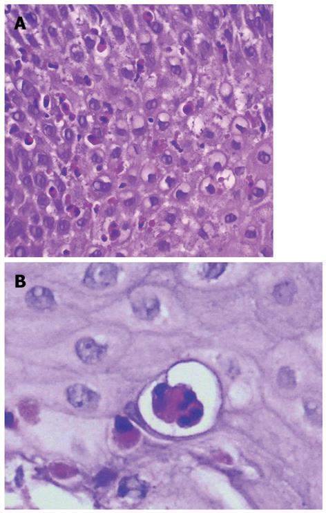
Figure 2 Histological findings in esophageal biopsy specimen.
A: Dense eosinophilic infiltrates; B: Microabscesses on esophageal microscopy.
- Citation: Caetano AC, Gonçalves R, Rolanda C. Eosinophilic esophagitis-endoscopic distinguishing findings. World J Gastroenterol 2012; 18(31): 4221-4223
- URL: https://www.wjgnet.com/1007-9327/full/v18/i31/4221.htm
- DOI: https://dx.doi.org/10.3748/wjg.v18.i31.4221


