Copyright
©2012 Baishideng Publishing Group Co.
World J Gastroenterol. Aug 21, 2012; 18(31): 4136-4144
Published online Aug 21, 2012. doi: 10.3748/wjg.v18.i31.4136
Published online Aug 21, 2012. doi: 10.3748/wjg.v18.i31.4136
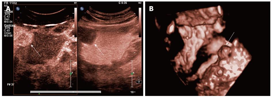
Figure 4 Double contrast-enhanced ultrasound imaging of gastric stromal tumor.
A: Two-dimensional double contrast-enhanced ultrasound (DCUS) images displayed a anechoic mass into the gastric cavity in oral contrast ultrasonography (right figure), from which we hardly judged whether it was cystic or solid lesion; but the intravenous contrast imaging (left figure) showed there was contrast agent enhancement in the focus of infection (arrow); B: Three-dimensional DCUS imaging displayed the tumor elevated to the gastric cavity (arrow).
- Citation: Shi H, Yu XH, Guo XZ, Guo Y, Zhang H, Qian B, Wei ZR, Li L, Wang XC, Kong ZX. Double contrast-enhanced two-dimensional and three-dimensional ultrasonography for evaluation of gastric lesions. World J Gastroenterol 2012; 18(31): 4136-4144
- URL: https://www.wjgnet.com/1007-9327/full/v18/i31/4136.htm
- DOI: https://dx.doi.org/10.3748/wjg.v18.i31.4136


