Copyright
©2012 Baishideng Publishing Group Co.
World J Gastroenterol. Aug 21, 2012; 18(31): 4102-4117
Published online Aug 21, 2012. doi: 10.3748/wjg.v18.i31.4102
Published online Aug 21, 2012. doi: 10.3748/wjg.v18.i31.4102
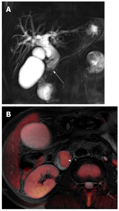
Figure 18 Ampullary carcinoma in a 63-year-old man.
A: Coronal image from thick-slab single-shot MRCP shows marked bile duct dilatation with abrupt narrowing (arrow) at the distal common bile duct; B: Fusion image of T2-weighted and diffusion-weighted imaging at b = 800 s/mm2 shows an ampullary mass with hyperintensity (arrowhead). MRCP: Magnetic resonance cholangiopancreatography.
- Citation: Lee NK, Kim S, Kim GH, Kim DU, Seo HI, Kim TU, Kang DH, Jang HJ. Diffusion-weighted imaging of biliopancreatic disorders: Correlation with conventional magnetic resonance imaging. World J Gastroenterol 2012; 18(31): 4102-4117
- URL: https://www.wjgnet.com/1007-9327/full/v18/i31/4102.htm
- DOI: https://dx.doi.org/10.3748/wjg.v18.i31.4102


