Copyright
©2012 Baishideng Publishing Group Co.
World J Gastroenterol. Aug 21, 2012; 18(31): 4102-4117
Published online Aug 21, 2012. doi: 10.3748/wjg.v18.i31.4102
Published online Aug 21, 2012. doi: 10.3748/wjg.v18.i31.4102
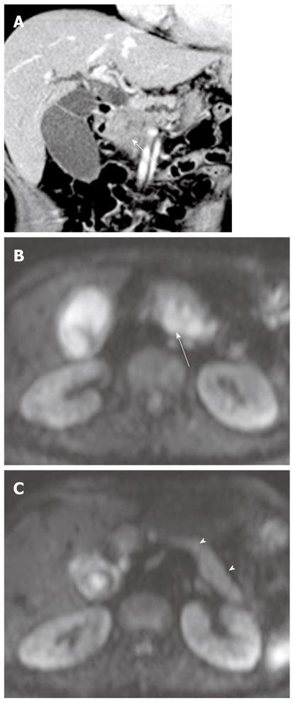
Figure 16 Pancreatic adenocarcinoma in a 50-year-old woman.
A: Coronal reformatted contrast-enhanced CT image shows a double duct sign secondary to a pancreatic head cancer (arrow); B: On DWI at b = 800 s/mm2, pancreatic adenocarcinoma (arrow) shows hyperintensity; C: DWI at b = 800 s/mm2 superior to (B) shows high signal intensity of the remaining pancreas (arrowheads) due to obstructive pancreatitis. However, the remaining pancreas is less hyperintense relative to pancreatic adenocarcinoma. The ADC value of pancreatic cancer (1.23 ± 0.32 mm2/s) is significantly lower than that of the remaining pancreas (1.85 ± 0.45 mm2/s). DWI: Diffusion-weighted magnetic resonance imaging; CT: Computed tomography; ADC: Apparent diffusion coefficient.
- Citation: Lee NK, Kim S, Kim GH, Kim DU, Seo HI, Kim TU, Kang DH, Jang HJ. Diffusion-weighted imaging of biliopancreatic disorders: Correlation with conventional magnetic resonance imaging. World J Gastroenterol 2012; 18(31): 4102-4117
- URL: https://www.wjgnet.com/1007-9327/full/v18/i31/4102.htm
- DOI: https://dx.doi.org/10.3748/wjg.v18.i31.4102


