Copyright
©2012 Baishideng Publishing Group Co.
World J Gastroenterol. Aug 21, 2012; 18(31): 4102-4117
Published online Aug 21, 2012. doi: 10.3748/wjg.v18.i31.4102
Published online Aug 21, 2012. doi: 10.3748/wjg.v18.i31.4102
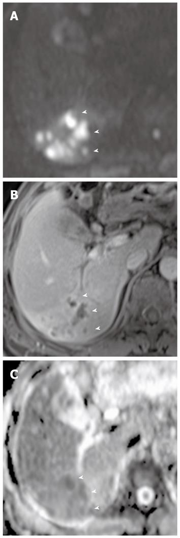
Figure 5 Liver abscesses complicating acute cholangitis in a 79-year-old man.
A: DWI at b = 1000 s/mm2 shows multiple liver abscesses with high signal intensity (arrowheads); B: Multiple abscesses with peripheral rim enhancement (arrowheads) are less conspicuous on contrast-enhanced fat-saturated T1-weighted images (B) than on DWI (A); C: On an ADC map, multiple abscesses appear as low signal intensity (arrowheads) due to restriction of diffusion. DWI: Diffusion-weighted magnetic resonance imaging; ADC: Apparent diffusion coefficient.
- Citation: Lee NK, Kim S, Kim GH, Kim DU, Seo HI, Kim TU, Kang DH, Jang HJ. Diffusion-weighted imaging of biliopancreatic disorders: Correlation with conventional magnetic resonance imaging. World J Gastroenterol 2012; 18(31): 4102-4117
- URL: https://www.wjgnet.com/1007-9327/full/v18/i31/4102.htm
- DOI: https://dx.doi.org/10.3748/wjg.v18.i31.4102


