Copyright
©2012 Baishideng Publishing Group Co.
World J Gastroenterol. Aug 21, 2012; 18(31): 4102-4117
Published online Aug 21, 2012. doi: 10.3748/wjg.v18.i31.4102
Published online Aug 21, 2012. doi: 10.3748/wjg.v18.i31.4102
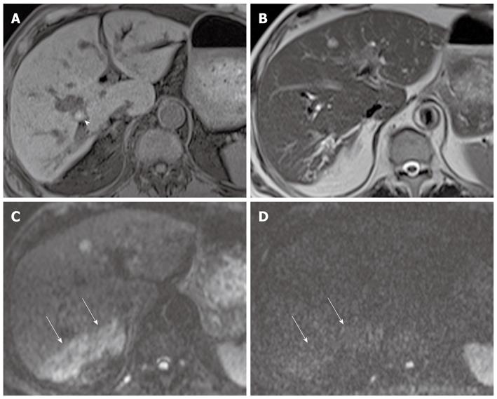
Figure 4 Parenchymal changes in acute cholangitis in a 76-year-old man.
A: Axial fat-saturated T1-weighted image shows a hyperintense stone (arrowhead) with upstream bile duct dilation; B: Axial T2-weighted rapid acquisition relaxation enhancement image shows mild bile duct dilatation in the right lobe of the liver, but no definite area of increased parenchymal signal intensity; C: DWI at b = 50 s/mm2 shows wedge-shaped areas of increased parenchymal signal intensity in segment 6 (arrows). Parenchymal changes are more conspicuous on black-blood images than on routine T2-weighted images; D: DWI at b = 800 s/mm2 shows that most areas of increased parenchymal signal intensity usually return to isointensity (arrows). Such a finding can be a clue for differentiating parenchymal changes due to cholangitis from abscesses. DWI: Diffusion-weighted magnetic resonance imaging.
- Citation: Lee NK, Kim S, Kim GH, Kim DU, Seo HI, Kim TU, Kang DH, Jang HJ. Diffusion-weighted imaging of biliopancreatic disorders: Correlation with conventional magnetic resonance imaging. World J Gastroenterol 2012; 18(31): 4102-4117
- URL: https://www.wjgnet.com/1007-9327/full/v18/i31/4102.htm
- DOI: https://dx.doi.org/10.3748/wjg.v18.i31.4102


