Copyright
©2012 Baishideng Publishing Group Co.
World J Gastroenterol. Aug 21, 2012; 18(31): 4086-4090
Published online Aug 21, 2012. doi: 10.3748/wjg.v18.i31.4086
Published online Aug 21, 2012. doi: 10.3748/wjg.v18.i31.4086
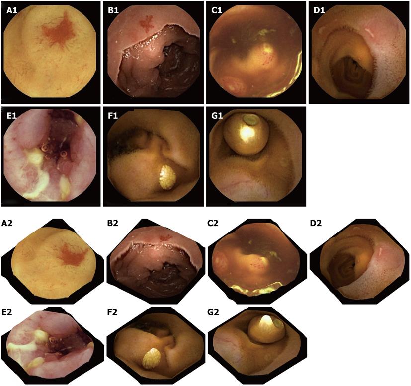
Figure 4 Two-dimensional capsule endoscopy images (upper panel) and three-dimensional representation of above structures is seen (lower panel).
A1, A2: P2 angioectasia; B1, B2: P1 angiectasia; C1, C2: Lymphoid hyperplasia with superficial ulceration; D1, D2: Aphthous ulcers; E1, E2: Serpiginous ulcers; F1, F2: Nodular lymphangiectasia; G1, G2: Another capsule endoscope still inside the small-bowel.
- Citation: Koulaouzidis A, Karargyris A. Three-dimensional image reconstruction in capsule endoscopy. World J Gastroenterol 2012; 18(31): 4086-4090
- URL: https://www.wjgnet.com/1007-9327/full/v18/i31/4086.htm
- DOI: https://dx.doi.org/10.3748/wjg.v18.i31.4086


