Copyright
©2012 Baishideng Publishing Group Co.
World J Gastroenterol. Aug 14, 2012; 18(30): 4064-4068
Published online Aug 14, 2012. doi: 10.3748/wjg.v18.i30.4064
Published online Aug 14, 2012. doi: 10.3748/wjg.v18.i30.4064
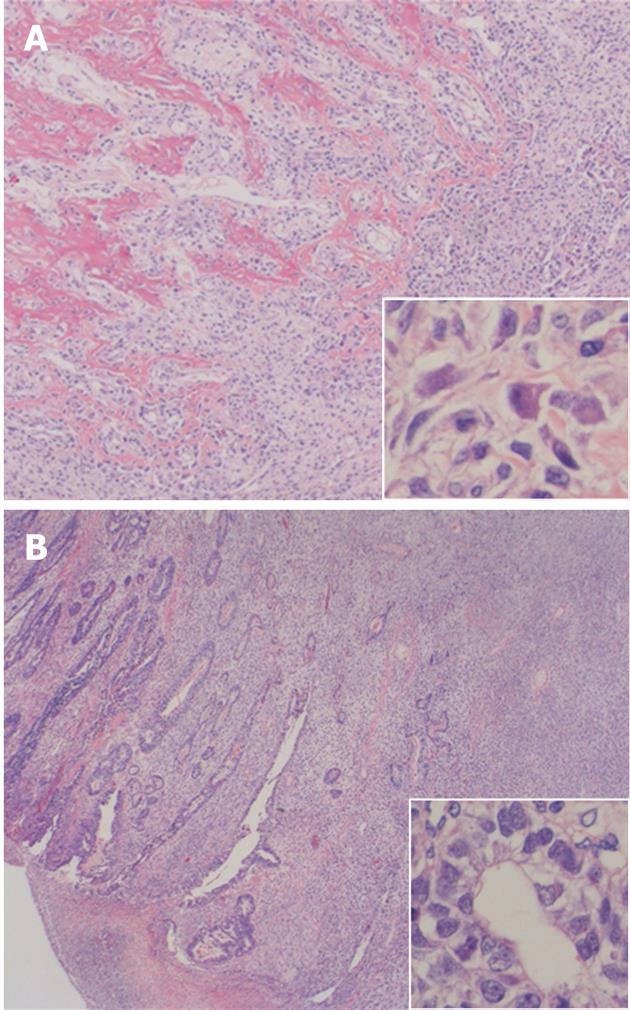
Figure 3 Representative microphotographs.
A: Hematoxylin and eosin stain of the tumor (4 ×), a large part of the tumor comprised spindle cells producing lace-like osteoid; B: Tubular adenocarcinoma coexisting in the tumor. High-power view of tumor cells (40 ×) is shown in the insets.
- Citation: Yoshida H, Tanaka N, Tochigi N, Suzuki Y. Rapidly deforming gastric carcinosarcoma with osteoblastic component: An autopsy case report. World J Gastroenterol 2012; 18(30): 4064-4068
- URL: https://www.wjgnet.com/1007-9327/full/v18/i30/4064.htm
- DOI: https://dx.doi.org/10.3748/wjg.v18.i30.4064


