Copyright
©2012 Baishideng Publishing Group Co.
World J Gastroenterol. Jan 21, 2012; 18(3): 291-294
Published online Jan 21, 2012. doi: 10.3748/wjg.v18.i3.291
Published online Jan 21, 2012. doi: 10.3748/wjg.v18.i3.291
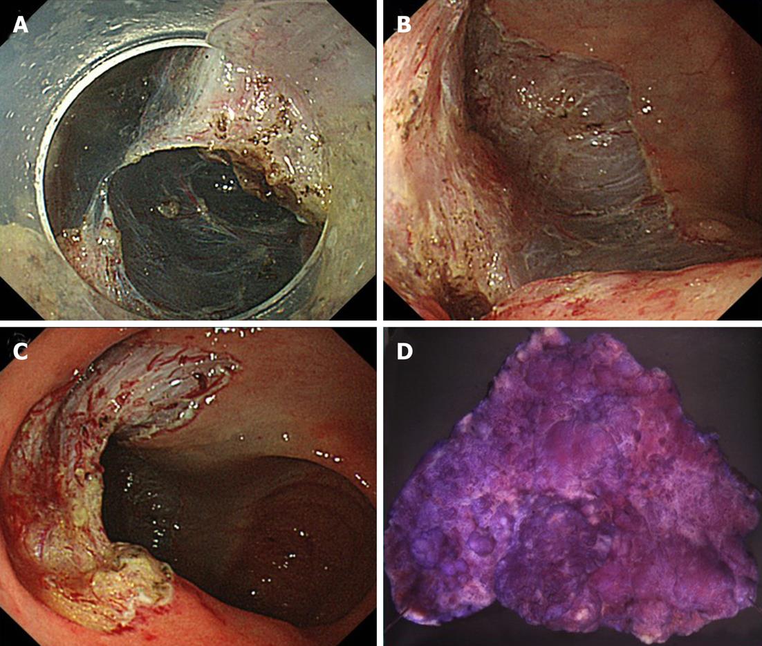
Figure 2 Procedure.
A: Endoscopic view through the distal attachment showing dissection with insulation-tip knife; B: Carefully check for bleeding throughout the ileocecal region; C: The ulcer bed of ileum after en-bloc endoscopic submucosal dissection; D: Stereomicroscopic view presenting the resected specimen, which pathology reported as a Is+IIa intramucosal cancer with tumor-free margins of 70 mm in diameter.
- Citation: Kishimoto G, Saito Y, Takisawa H, Suzuki H, Sakamoto T, Nakajima T, Matsuda T. Endoscopic submucosal dissection for large laterally spreading tumors involving the ileocecal valve and terminal ileum. World J Gastroenterol 2012; 18(3): 291-294
- URL: https://www.wjgnet.com/1007-9327/full/v18/i3/291.htm
- DOI: https://dx.doi.org/10.3748/wjg.v18.i3.291


