Copyright
©2012 Baishideng Publishing Group Co.
World J Gastroenterol. Jan 21, 2012; 18(3): 291-294
Published online Jan 21, 2012. doi: 10.3748/wjg.v18.i3.291
Published online Jan 21, 2012. doi: 10.3748/wjg.v18.i3.291
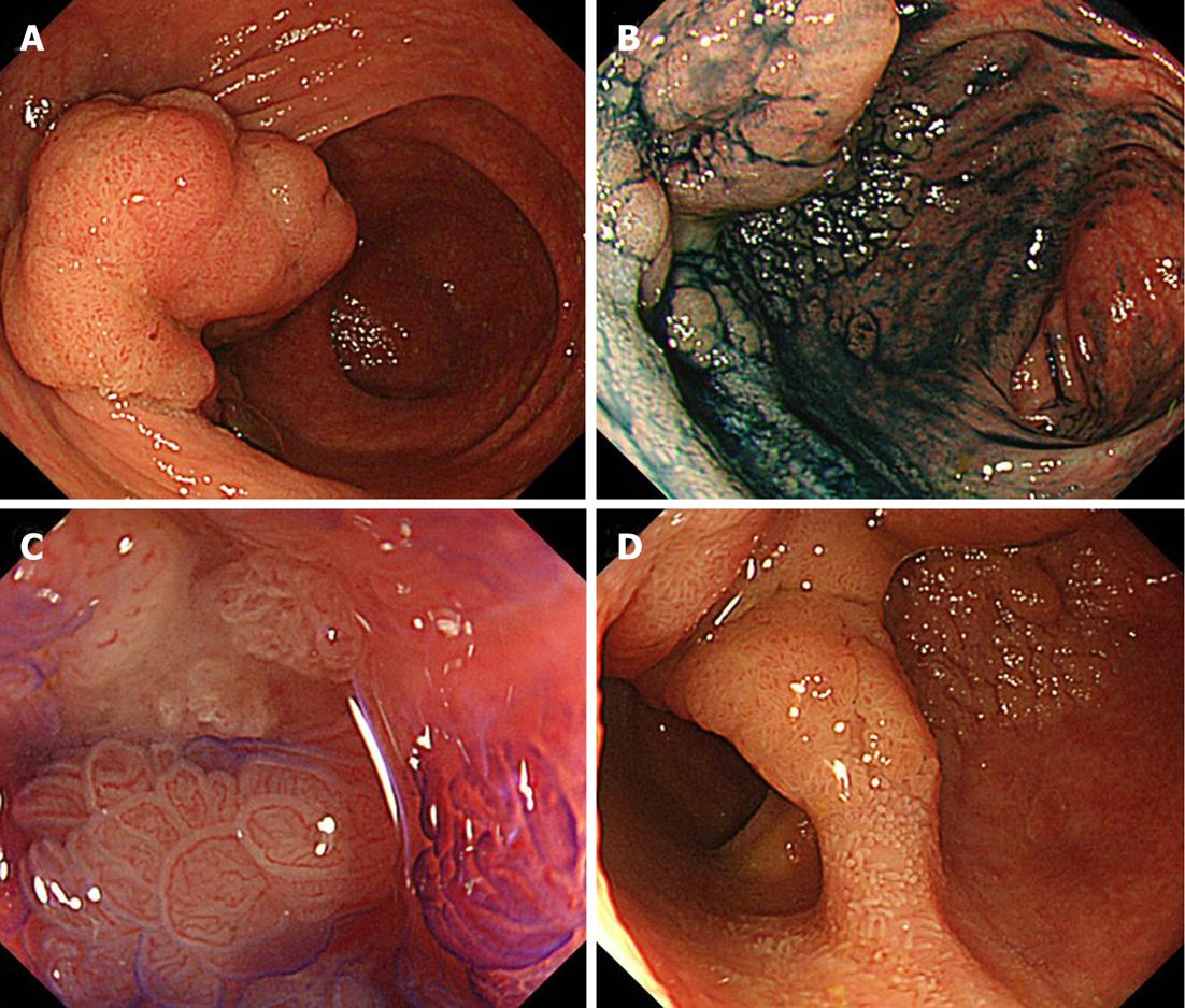
Figure 1 Pre-treatment endoscopic evaluation.
A: Close view of the cecum revealed a 70 mmIs +IIa, LST granular type (LST-G) lesion; B: Clearly delineated margin of the LST-G lesion after 0.4% indigo-carmine dye spraying; C: Magnification view of the Is component of the Is +IIa (LST-G); D: Spreading confirmation of the tumor through the ileocecal valve to the terminal ileum.
- Citation: Kishimoto G, Saito Y, Takisawa H, Suzuki H, Sakamoto T, Nakajima T, Matsuda T. Endoscopic submucosal dissection for large laterally spreading tumors involving the ileocecal valve and terminal ileum. World J Gastroenterol 2012; 18(3): 291-294
- URL: https://www.wjgnet.com/1007-9327/full/v18/i3/291.htm
- DOI: https://dx.doi.org/10.3748/wjg.v18.i3.291


