Copyright
©2012 Baishideng Publishing Group Co.
World J Gastroenterol. Jul 28, 2012; 18(28): 3761-3764
Published online Jul 28, 2012. doi: 10.3748/wjg.v18.i28.3761
Published online Jul 28, 2012. doi: 10.3748/wjg.v18.i28.3761
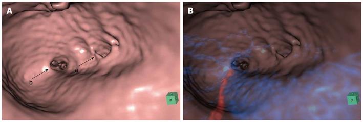
Figure 3 Virtual cholangioscopy.
A: Virtual cholangioscopy demonstrating a relative stricture of the orifice in the posterior branch (a) and no stricture in the anterior branch (b); B: Composite virtual cholangioscopy demonstrating no relationship between the membrane-like stricture and the surrounding blood vessels. Red indicates the hepatic artery and blue denotes the portal vein.
- Citation: Tsuchida A, Nagakawa Y, Kasuya K, Kyo B, Ikeda T, Suzuki Y, Aoki T, Itoi T. Computed tomography virtual endoscopy with angiographic imaging for the treatment of type IV-A choledochal cyst. World J Gastroenterol 2012; 18(28): 3761-3764
- URL: https://www.wjgnet.com/1007-9327/full/v18/i28/3761.htm
- DOI: https://dx.doi.org/10.3748/wjg.v18.i28.3761


