Copyright
©2012 Baishideng Publishing Group Co.
World J Gastroenterol. Jul 28, 2012; 18(28): 3696-3704
Published online Jul 28, 2012. doi: 10.3748/wjg.v18.i28.3696
Published online Jul 28, 2012. doi: 10.3748/wjg.v18.i28.3696
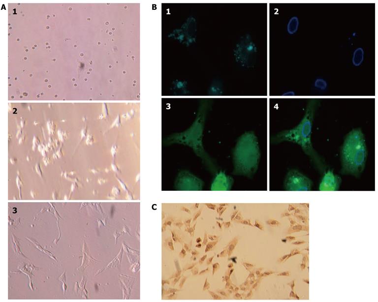
Figure 2 Isolation, culture, and identification of primary rat hepatic stellate cells.
A: Primary rat hepatic stellate cells (HSCs) on days 1, 2, and 5 after isolation and plated in culture; B: HSCs isolated from Sprague-Dawley rats showed characteristic multiple lipid droplets with a rapid fade of the blue-green autofluorescence at 328 nm. 1: Lipid droplets; 2: Cell nucleus; 3: Cell body. 4: Merged images; C: HSC culture after the first passage and stained with anti-α-smooth muscle actin antibody.
- Citation: Zhang SC, Zheng YH, Yu PP, Min TH, Yu FX, Ye C, Xie YK, Zhang QY. Lentiviral vector-mediated down-regulation of IL-17A receptor in hepatic stellate cells results in decreased secretion of IL-6. World J Gastroenterol 2012; 18(28): 3696-3704
- URL: https://www.wjgnet.com/1007-9327/full/v18/i28/3696.htm
- DOI: https://dx.doi.org/10.3748/wjg.v18.i28.3696


