Copyright
©2012 Baishideng Publishing Group Co.
World J Gastroenterol. Jul 28, 2012; 18(28): 3681-3695
Published online Jul 28, 2012. doi: 10.3748/wjg.v18.i28.3681
Published online Jul 28, 2012. doi: 10.3748/wjg.v18.i28.3681
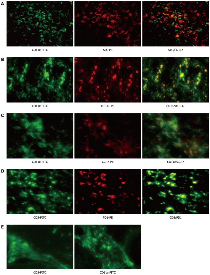
Figure 4 Localization of CD11c and programmed death 1 in liver tissue from acute hepatitis B and chronic hepatitis B virus tolerance patients.
A-C: Colocalization of CD11c with SLC, MIP3α and CCR7 by immunofluorescence double staining in liver biopsy specimens of acute hepatitis B (AHB) patients. CD11c (green) is co-localized with SLC (red) (A), MIP3α (red) (B) and CCR7 (red) (C). The 2-color merged panels were shown with colocalization visible in yellow. Original magnification × 200; D: Colocalization of CD8 with programmed death 1 (PD1) by immunofluorescence double staining in liver biopsy specimens of AHB patients. CD8 (green) is co-localized with PD1 (red). Original magnification × 200; E: Inflammatory cells, such as CD8 and CD11c positive cells, can be observed mainly in fibrous septa in the liver tissue of chronic hepatitis B virus tolerance patients. FITC: Fluorescein isothiocyanate; PE: Phycoerythrin; SLC: Secondary lymphoid tissue chemokine; MIP3α: Macrophage inflammatory protein 3 α; CCR: Chemokine (C-C) receptor.
- Citation: Zhang WJ, Xie HY, Duan X, Wan YL, Peng CH, Shi SH, Su R, Zheng ZH, Pan LL, Zhou L, Zheng SS. Study of human B7 homolog 1 expression in patients with hepatitis B virus infection. World J Gastroenterol 2012; 18(28): 3681-3695
- URL: https://www.wjgnet.com/1007-9327/full/v18/i28/3681.htm
- DOI: https://dx.doi.org/10.3748/wjg.v18.i28.3681


