Copyright
©2012 Baishideng Publishing Group Co.
World J Gastroenterol. Jul 21, 2012; 18(27): 3527-3536
Published online Jul 21, 2012. doi: 10.3748/wjg.v18.i27.3527
Published online Jul 21, 2012. doi: 10.3748/wjg.v18.i27.3527
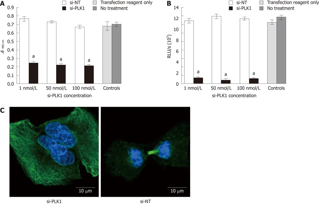
Figure 2 Reduction of cell proliferation by 3-(4,5-dimethylthiazol-2-yl)-5-(3-carboxymethoxyphenyl)-2(4-sulfophenyl)-2H-tetrazolium assay and bromodeoxyuridine assay after silencing of polo-like kinase 1, and failure of mitosis after knockdown of polo-like kinase 1.
A: Knockdown of polo-like kinase 1 (PLK1) reduced cell proliferation in Huh-7 cells in the 3-(4,5-dimethylthiazol-2-yl)-5-(3-carboxymethoxyphenyl)-2(4-sulfophenyl)-2H-tetrazolium (MTS) cell proliferation assay by a mean of 65% compared with short interfering non-targeting (si-NT) or controls; B: Knockdown of PLK1 reduced cell proliferation in the bromodeoxyuridine cell proliferation assay in Huh7 cells by a mean of 93% with 50 nmol/L short-interfering PLK1 (si-PLK1) compared with si-NT or controls; C: Confocal fluorescence images show the si-PLK1 transfected Huh-7 cells (left panel) were binucleated, depicting failure in completing mitosis due to the lack of a functional spindle assembly. The right panel shows a functional spindle assembly in Huh-7 cells transfected with si-NT. Huh-7 cells were processed for confocal imaging after 24 h of transfection either with 50 nmol/L si-PLK1 or si-NT; α-tubulins were stained with fluorescein isothiocyanate-conjugated antibody and nuclei were counterstained with 4',6-diamidino-2-phenylindole. Data are shown as mean ± SE, using the Student t-test (aP < 0.05).
- Citation: Mok WC, Wasser S, Tan T, Lim SG. Polo-like kinase 1, a new therapeutic target in hepatocellular carcinoma. World J Gastroenterol 2012; 18(27): 3527-3536
- URL: https://www.wjgnet.com/1007-9327/full/v18/i27/3527.htm
- DOI: https://dx.doi.org/10.3748/wjg.v18.i27.3527


