Copyright
©2012 Baishideng Publishing Group Co.
World J Gastroenterol. Jul 7, 2012; 18(25): 3322-3326
Published online Jul 7, 2012. doi: 10.3748/wjg.v18.i25.3322
Published online Jul 7, 2012. doi: 10.3748/wjg.v18.i25.3322
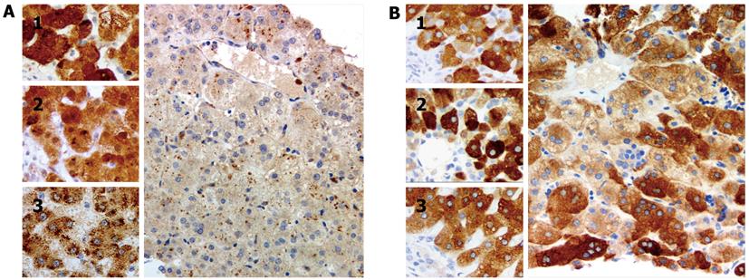
Figure 2 Photomicrographs of liver immunostained for enzymes that subserve bile-acid amidation.
A: Main image: Core needle biopsy specimen, patient liver (age 57 d); inset, control livers (1, infant, matched for age with patient, with cholestasis and extrahepatic biliary atresia; 2, infant, matched for age with patient, with intrahepatic cholestasis in setting of absence of bile salt export pump (BSEP) expression; and 3, adult without cholestasis). The patient liver lacks immunohistochemically demonstrable bile acid-CoA: amino acid N-acyltransferase (BAAT). Granular cytoplasmic marking, suggestive of peroxisomal location, is found in (3); marking is more diffuse in the livers of age-matched infants; B: Tissue specimens as in (A). Diffuse cytoplasmic marking for cholate-CoA ligase (CCL) is seen in patient liver (main image) and in control livers (1, infant, matched for age with patient, with cholestasis and extrahepatic biliary atresia; 2, infant, matched for age with patient, with intrahepatic cholestasis in setting of absence of BSEP expression; and 3, adult without cholestasis). A, anti-BAAT antibody, and B, anti-CCL antibody (see principal text for sources); in all images, reaction development with EnVision proprietary technique (Dako UK, Ely, United Kingdom), haematoxylin counterstain, original magnification 200 ×.
- Citation: Hadžić N, Bull LN, Clayton PT, Knisely A. Diagnosis in bile acid-CoA: Amino acid N-acyltransferase deficiency. World J Gastroenterol 2012; 18(25): 3322-3326
- URL: https://www.wjgnet.com/1007-9327/full/v18/i25/3322.htm
- DOI: https://dx.doi.org/10.3748/wjg.v18.i25.3322


