Copyright
©2012 Baishideng Publishing Group Co.
World J Gastroenterol. Jun 14, 2012; 18(22): 2872-2876
Published online Jun 14, 2012. doi: 10.3748/wjg.v18.i22.2872
Published online Jun 14, 2012. doi: 10.3748/wjg.v18.i22.2872
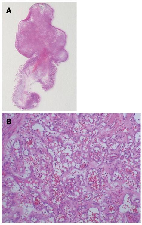
Figure 4 Pathological specimen.
A: A magnified pathological image of the resected specimen (10 ×); B: A magnified pathological image of the central part of the 20 mm × 12 mm tumor, which shows proliferation of various sized capillary lumens (100 ×).
- Citation: Nishiyama N, Mori H, Kobara H, Fujihara S, Nomura T, Kobayashi M, Masaki T. Bleeding duodenal hemangioma: Morphological changes and endoscopic mucosal resection. World J Gastroenterol 2012; 18(22): 2872-2876
- URL: https://www.wjgnet.com/1007-9327/full/v18/i22/2872.htm
- DOI: https://dx.doi.org/10.3748/wjg.v18.i22.2872


