Copyright
©2012 Baishideng Publishing Group Co.
World J Gastroenterol. Jun 14, 2012; 18(22): 2844-2849
Published online Jun 14, 2012. doi: 10.3748/wjg.v18.i22.2844
Published online Jun 14, 2012. doi: 10.3748/wjg.v18.i22.2844
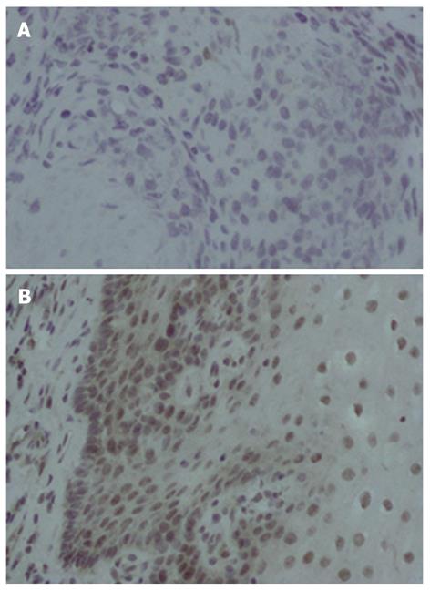
Figure 3 Immunohistochemistry analysis of X chromosome-linked inhibitor of apoptosis-associated factor 1 in esophageal cancer tissue and adjacent tissue.
Esophageal cancer and adjacent normal tissue samples were immunohistologically analyzed with anti-X chromosome-linked inhibitor of apoptosis-associated factor 1 (XAF1) (1:200 dilution; × 400). A: XAF1 was not detected in esophageal cancer tissue; B: XAF1 was localized in the nucleus and cytoplasm in adjacent normal esophageal tissue.
- Citation: Chen XY, He QY, Guo MZ. XAF1 is frequently methylated in human esophageal cancer. World J Gastroenterol 2012; 18(22): 2844-2849
- URL: https://www.wjgnet.com/1007-9327/full/v18/i22/2844.htm
- DOI: https://dx.doi.org/10.3748/wjg.v18.i22.2844


