Copyright
©2012 Baishideng Publishing Group Co.
World J Gastroenterol. Jun 14, 2012; 18(22): 2798-2804
Published online Jun 14, 2012. doi: 10.3748/wjg.v18.i22.2798
Published online Jun 14, 2012. doi: 10.3748/wjg.v18.i22.2798
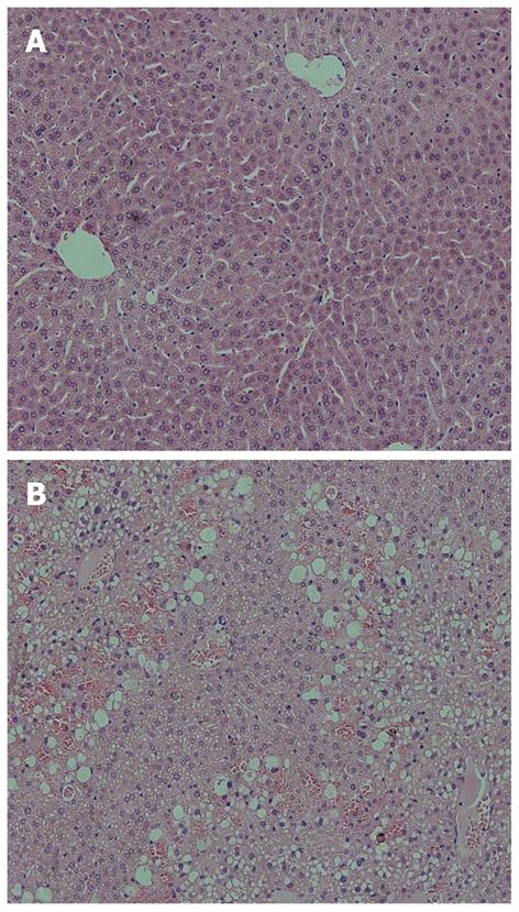
Figure 4 Histopathologic analysis of liver after lethal compared to sublethal acetaminophen dosing.
Hematoxylin and eosin liver tissue staining at 12 h of (A) sublethally (150 mg/kg) acetaminophen poisoned mice, with no signs of centrilobular inflammation or necrosis; and (B) lethally (500 mg/kg) acetaminophen poisoned mice with extensive centrilobular necrosis, enlarged hepatocytes, and highly vacuolated cytoplasm.
- Citation: Ward J, Bala S, Petrasek J, Szabo G. Plasma microRNA profiles distinguish lethal injury in acetaminophen toxicity: A research study. World J Gastroenterol 2012; 18(22): 2798-2804
- URL: https://www.wjgnet.com/1007-9327/full/v18/i22/2798.htm
- DOI: https://dx.doi.org/10.3748/wjg.v18.i22.2798


