Copyright
©2012 Baishideng Publishing Group Co.
World J Gastroenterol. Jun 14, 2012; 18(22): 2775-2783
Published online Jun 14, 2012. doi: 10.3748/wjg.v18.i22.2775
Published online Jun 14, 2012. doi: 10.3748/wjg.v18.i22.2775
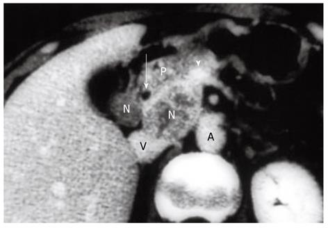
Figure 2 Computed tomography revealing enlarged peripancreatic nodes with heterogeneous contrast enhancement (N).
Note that the posterior superior pancreaticoduodenal node (N, left) and the retroportal node (N, right) are adhered to the head of the pancreas (P). The latter node shifts the portal vein (arrowhead) in a right-upper direction. Histological examination confirmed metastases in 10 regional nodes, some of which had invaded the pancreatic parenchyma. This patient remains well with no evidence of disease at 15 years after a pancreaticoduodenectomy combined with wedge resection of the gallbladder fossa. The arrow indicates common bile duct; V: Inferior vena cava; A: Aorta.
- Citation: Shirai Y, Wakai T, Sakata J, Hatakeyama K. Regional lymphadenectomy for gallbladder cancer: Rational extent, technical details, and patient outcomes. World J Gastroenterol 2012; 18(22): 2775-2783
- URL: https://www.wjgnet.com/1007-9327/full/v18/i22/2775.htm
- DOI: https://dx.doi.org/10.3748/wjg.v18.i22.2775


