Copyright
©2012 Baishideng Publishing Group Co.
World J Gastroenterol. Jun 14, 2012; 18(22): 2767-2774
Published online Jun 14, 2012. doi: 10.3748/wjg.v18.i22.2767
Published online Jun 14, 2012. doi: 10.3748/wjg.v18.i22.2767
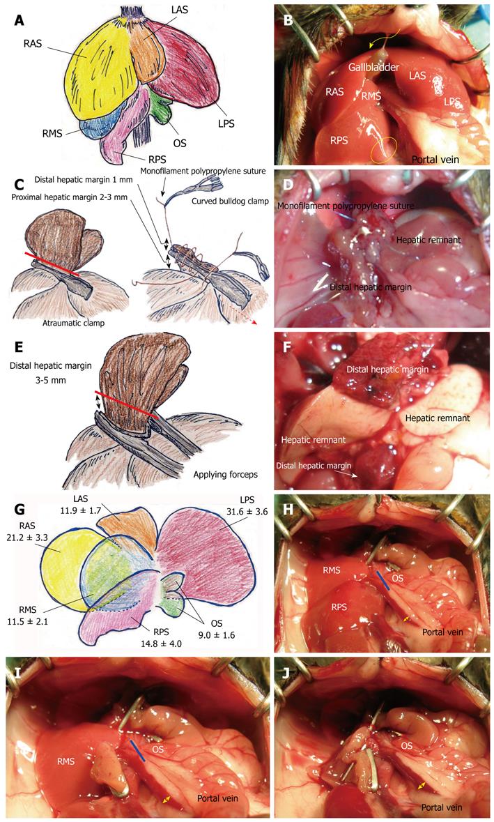
Figure 1 Hepatic segments and hepatectomy by suture or clip techniques.
A: The murine liver is divided into six segments (RAS, RMS, RPS, LAS, LPS, and OS); B: The gallbladder is detected between the RAS and LAS; C: Atraumatic clamp was placed on hepatic segment, with optimal proximal hepatic margin of 2-3 mm from division of each segment. Clamped segment was sharply cut with 1 mm distal hepatic margin (red line). Sutures not involving the clamp were placed bilaterally. Bilateral sutures were ligated, and each suture was grasped with a curved bulldog clamp. By using suture of left-side ligation, cut surface of liver was sutured from left side to right side with loose continuous suture that involved a clamp, and the last suture reached the right side; D: Clamp was removed (dotted red arrow) and hepatic margin was immediately treated by tightening the continuous suture. The thread of tightened continuous suture was ligated with stay suture of right side without over-tightening. Continuous suture was made from right side to left side. The last suture was ligated with stay suture of left side without over-tightening; E: The relevant segment was cut with a 3-5 mm distal hepatic margin (red line); F: Ischemic change of distal hepatic margin at 24 h after 80% hepatectomy was shown. Although liver remnants after massive hepatectomy showed color change due to vacuolization, distal hepatic margins clearly showed necrotic changes. Color change between liver remnant and distal hepatic margin was more enhanced at autopsy; G: Percentages of total volume of each segment were 21.2% ± 3.3% for RAS, 11.5% ± 2.1% for RMS, 14.8% ± 4.0% for RPS, 11.9% ± 1.7% for LAS, 31.6% ± 3.6% for LPS, and 9.0% ± 1.6% for OS; H: Traditional 2/3 hepatectomy of RMS + RPS + OS (35.4% ± 4.0%) is shown. Additional resection of OS (blue line) will make a 75% hepatectomy (RMS + RPS, 26.4% ± 3.8 %); I: Hepatic remnant in 80% hepatectomy was RMS + OS (20.6% ± 2.6%). Additional resection of OS (blue line) will make an RMS-remnant 90% hepatectomy (RMS, 14.8% ± 4.0%); J: OS-remnant 90% hepatectomy is shown (OS, 9.0% ± 1.6%). Dilation of portal vein due to portal hypertension is confirmed in reverse proportion to volume of hepatic remnant (yellow arrows). LAS: Left anterior segment; LPS: Left posterior segment; OS: Omental segment; RAS: Right anterior segment; RMS: Right middle segment; RPS: Right posterior segment.
- Citation: Hori T, Ohashi N, Chen F, Baine AMT, Gardner LB, Hata T, Uemoto S, Nguyen JH. Simple and reproducible hepatectomy in the mouse using the clip technique. World J Gastroenterol 2012; 18(22): 2767-2774
- URL: https://www.wjgnet.com/1007-9327/full/v18/i22/2767.htm
- DOI: https://dx.doi.org/10.3748/wjg.v18.i22.2767


