Copyright
©2012 Baishideng Publishing Group Co.
World J Gastroenterol. May 28, 2012; 18(20): 2452-2461
Published online May 28, 2012. doi: 10.3748/wjg.v18.i20.2452
Published online May 28, 2012. doi: 10.3748/wjg.v18.i20.2452
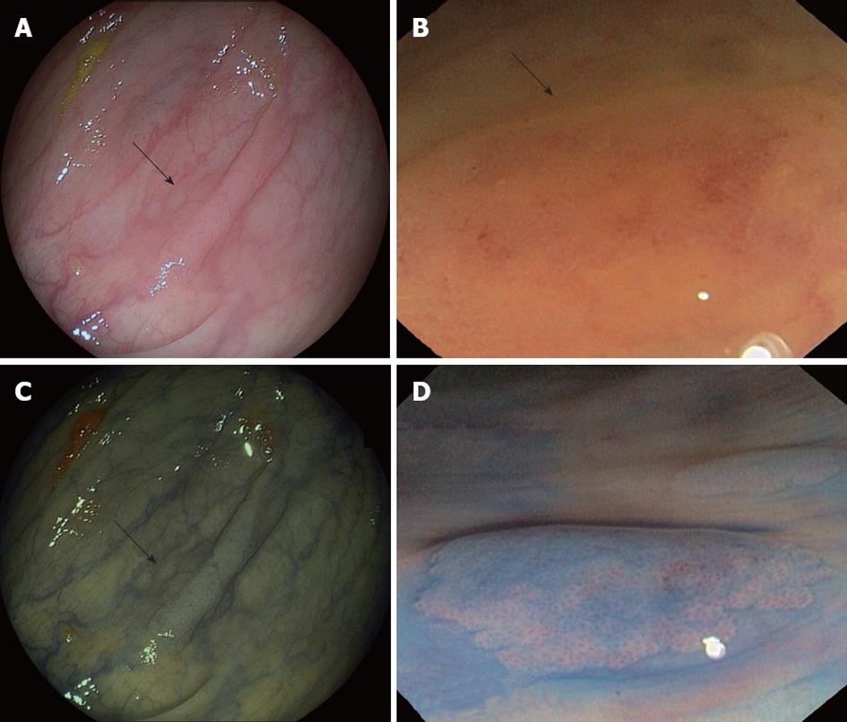
Figure 3 Endoscopic appearance of serrated polyps.
A and B: Sessile serrated adenoma (SSA) (arrows) as flat polyp on conventional optical colonoscopy; C: Narrow-band imaging appearance of polyp (arrow) seen in panel A; D: Chromoendoscopy image of SSA revealing Kudo II pattern (Images courtesy of Dr. Adolfo Parra, Hospital Central de Asturias, Oviedo, Spain).
- Citation: Guarinos C, Sánchez-Fortún C, Rodríguez-Soler M, Alenda C, Payá A, Jover R. Serrated polyposis syndrome: Molecular, pathological and clinical aspects. World J Gastroenterol 2012; 18(20): 2452-2461
- URL: https://www.wjgnet.com/1007-9327/full/v18/i20/2452.htm
- DOI: https://dx.doi.org/10.3748/wjg.v18.i20.2452


