Copyright
©2012 Baishideng Publishing Group Co.
World J Gastroenterol. May 21, 2012; 18(19): 2320-2333
Published online May 21, 2012. doi: 10.3748/wjg.v18.i19.2320
Published online May 21, 2012. doi: 10.3748/wjg.v18.i19.2320
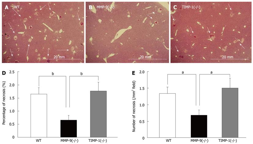
Figure 5 Histological analysis in each strain.
Histological analysis between wild-type (WT, n = 15) (A), matrix metalloproteinase (MMP)-9(-/-) (n = 15) (B), and tissue inhibitors of metalloproteinases (TIMP)-1 (-/-) mice (n = 14) (C) at 6 h after 80%-partial hepatectomy (PH). Representative images of remnant liver histology at 6 h after 80%-PH in WT, MMP-9(-/-), and TIMP-1(-/-) mice. Foci of hemorrhage and necrosis are denoted by white arrows. The percentage of the necrotic area (D) and the number of necrotic foci per mm2 (E) in remnant livers of study mice are shown. Significantly smaller and fewer necrotic foci in the remnant liver were observed in MMP-9(-/-) mice compared with WT and TIMP-1(-/-) mice (aP < 0.05, bP < 0.01).
- Citation: Ohashi N, Hori T, Chen F, Jermanus S, Eckman CB, Nakao A, Uemoto S, Nguyen JH. Matrix metalloproteinase-9 contributes to parenchymal hemorrhage and necrosis in the remnant liver after extended hepatectomy in mice. World J Gastroenterol 2012; 18(19): 2320-2333
- URL: https://www.wjgnet.com/1007-9327/full/v18/i19/2320.htm
- DOI: https://dx.doi.org/10.3748/wjg.v18.i19.2320


