Copyright
©2012 Baishideng Publishing Group Co.
World J Gastroenterol. May 21, 2012; 18(19): 2300-2308
Published online May 21, 2012. doi: 10.3748/wjg.v18.i19.2300
Published online May 21, 2012. doi: 10.3748/wjg.v18.i19.2300
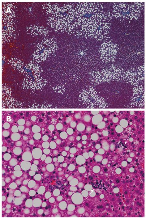
Figure 1 Liver histology of rats fed a high-fructose diet for 5 wk.
A: Hepatic steatosis, mainly distributed in zone 1, is observed (azan stain, × 40); B: Both macrovesicular and microvesicular steatosis are evident as well as scattered necroinflammatory foci (arrows) (hematoxylin and eosin stain, × 200).
- Citation: Takahashi Y, Soejima Y, Fukusato T. Animal models of nonalcoholic fatty liver disease/nonalcoholic steatohepatitis. World J Gastroenterol 2012; 18(19): 2300-2308
- URL: https://www.wjgnet.com/1007-9327/full/v18/i19/2300.htm
- DOI: https://dx.doi.org/10.3748/wjg.v18.i19.2300


