Copyright
©2012 Baishideng Publishing Group Co.
World J Gastroenterol. May 7, 2012; 18(17): 2140-2144
Published online May 7, 2012. doi: 10.3748/wjg.v18.i17.2140
Published online May 7, 2012. doi: 10.3748/wjg.v18.i17.2140
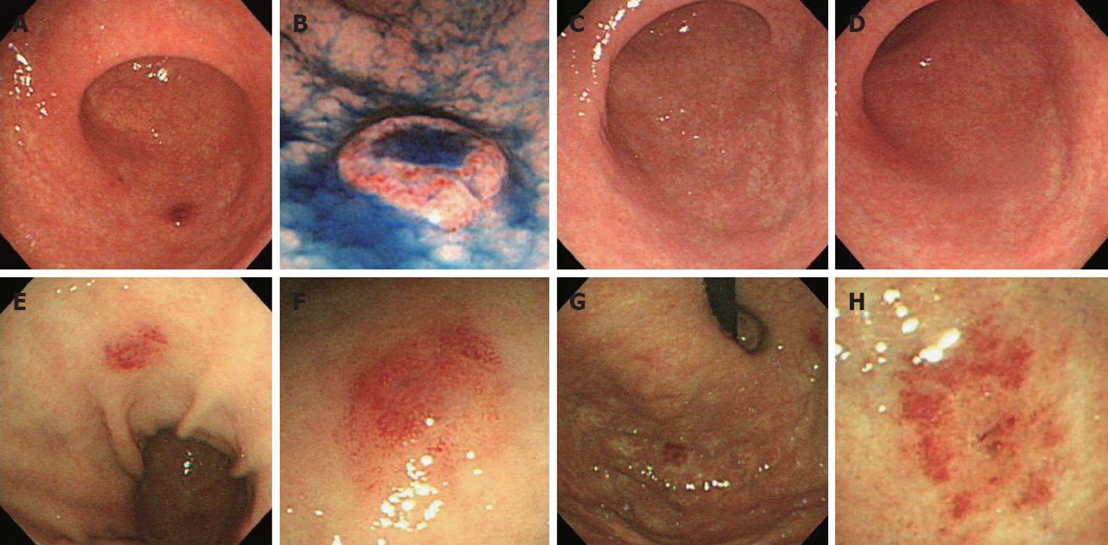
Figure 1 Gastroendoscopy revealed an erythematous dish-like elevated lesion in the greater curvature of the lower body at check-up (A and B), and at one (C) and six months later (D); Twelve months later, endoscopy revealed a similar lesion in the anterior wall of the middlebody (E and F), and an erythematous lesion in the fornix (G and H).
- Citation: Terai T, Sugimoto M, Uozaki H, Kitagawa T, Kinoshita M, Baba S, Yamada T, Osawa S, Sugimoto K. Lymphomatoidgastropathy mimicking extranodal NK/T cell lymphoma, nasal type: A case report. World J Gastroenterol 2012; 18(17): 2140-2144
- URL: https://www.wjgnet.com/1007-9327/full/v18/i17/2140.htm
- DOI: https://dx.doi.org/10.3748/wjg.v18.i17.2140


