Copyright
©2012 Baishideng Publishing Group Co.
World J Gastroenterol. May 7, 2012; 18(17): 2092-2098
Published online May 7, 2012. doi: 10.3748/wjg.v18.i17.2092
Published online May 7, 2012. doi: 10.3748/wjg.v18.i17.2092
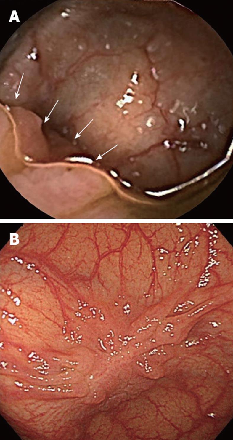
Figure 2 Example of the contribution of dimethicone to improved mucosal visualization by the colon capsule.
A: A lesion (arrow) is clearly observed on the transverse colon by colon capsule endoscopy. Dimethicone worked to join numerous microbubbles to form groups of large bubbles, resulting in better mucosal visualization; B: Colonoscopy image of the lesion post-capsule procedure. The lesion was diagnosed as a laterally spreading tumor. Endoscopic submucosal dissection was performed, and intramucosal cancer consisting of well differentiated adenocarcinoma was identified on the resected specimen.
- Citation: Kakugawa Y, Saito Y, Saito S, Watanabe K, Ohmiya N, Murano M, Oka S, Arakawa T, Goto H, Higuchi K, Tanaka S, Ishikawa H, Tajiri H. New reduced volume preparation regimen in colon capsule endoscopy. World J Gastroenterol 2012; 18(17): 2092-2098
- URL: https://www.wjgnet.com/1007-9327/full/v18/i17/2092.htm
- DOI: https://dx.doi.org/10.3748/wjg.v18.i17.2092


