Copyright
©2012 Baishideng Publishing Group Co.
World J Gastroenterol. May 7, 2012; 18(17): 2026-2034
Published online May 7, 2012. doi: 10.3748/wjg.v18.i17.2026
Published online May 7, 2012. doi: 10.3748/wjg.v18.i17.2026
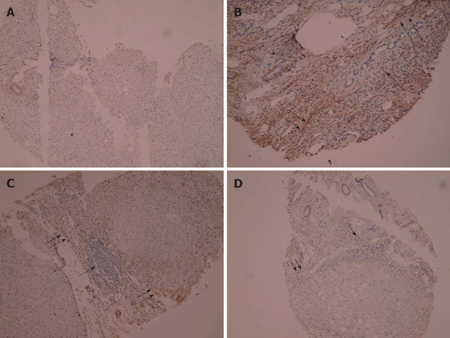
Figure 5 Immunohistochemical staining of α-smooth muscle actin.
A: Section of liver from controls showing negative staining (PAP × 125); B: Section of liver from liver fibrosis patients before treatment showing dense fibrotic reaction with strong positivity for α-smooth muscle actin (α-SMA) ↑↑ and dense mononuclear cellular infiltration ↑ (PAP × 225); C: Section of liver from conventional group after treatment showing dense mononuclear inflammatory infiltration ↑ and moderate positivity for α-SMA in fibrotic lesion↑↑ (PAP × 125); D: Section of liver from AHM group after treatment showing moderate mononuclear cellular infiltration ↑ surrounded by minimal fibrotic reaction stained mildly with α-SMA ↑↑ (PAP × 125).
- Citation: Hegazy SK, El-Bedewy M, Yagi A. Antifibrotic effect of aloe vera in viral infection-induced hepatic periportal fibrosis. World J Gastroenterol 2012; 18(17): 2026-2034
- URL: https://www.wjgnet.com/1007-9327/full/v18/i17/2026.htm
- DOI: https://dx.doi.org/10.3748/wjg.v18.i17.2026


