Copyright
©2012 Baishideng Publishing Group Co.
World J Gastroenterol. Apr 28, 2012; 18(16): 1991-1995
Published online Apr 28, 2012. doi: 10.3748/wjg.v18.i16.1991
Published online Apr 28, 2012. doi: 10.3748/wjg.v18.i16.1991
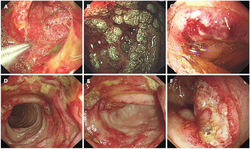
Figure 1 The endoscopy examination revealed intestinal damage.
A-C are gastroscopic images, which show significant damage to the duodenum. A: Diffuse redness, swelling, hemorrhage and petechiae in the mucosa; B: Distortion and proliferation of the duplicature (narrow-band image); C: Ulcer; D-F: Colonoscopic images and demonstrate significant damage to the terminal ileum; D: Mucosal hemorrhage and petechiae; E, F: Ulcers.
- Citation: Chen XL, Tian H, Li JZ, Tao J, Tang H, Li Y, Wu B. Paroxysmal drastic abdominal pain with tardive cutaneous lesions presenting in Henoch-Schönlein purpura. World J Gastroenterol 2012; 18(16): 1991-1995
- URL: https://www.wjgnet.com/1007-9327/full/v18/i16/1991.htm
- DOI: https://dx.doi.org/10.3748/wjg.v18.i16.1991


