Copyright
©2012 Baishideng Publishing Group Co.
World J Gastroenterol. Apr 28, 2012; 18(16): 1875-1883
Published online Apr 28, 2012. doi: 10.3748/wjg.v18.i16.1875
Published online Apr 28, 2012. doi: 10.3748/wjg.v18.i16.1875
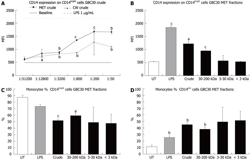
Figure 1 CD14 expression on mononuclear phagocytes.
Mononuclear phagocytes present in 3-d peripheral blood mononuclear cell cultures exposed to either the Ganeden Bacillus coagulans 30 (GBC30) metabolites (MET), cell wall-enriched (CW), or MET fractions, were identified using electronic gating of the flow cytometry data by gating on forward scatter/side scatter followed by gating for CD14 positivity. A comparison was made between cells that were untreated (UT), exposed to lipopolysaccharide (LPS) or to the different GBC30 fractions. A: Comparison of CD14 mean fluorescence intensity showed a dose-dependent increase in CD14 expression in cells treated with crude MET. A milder increase was seen for cells treated with crude CW. The baseline indicates CD14 expression on untreated cells; B: The increase in CD14 expression was primarily caused by high molecular weight compounds present in MET; C: The percent of CD14bright cells in the mononuclear phagocyte population was decreased by all fractions of MET; D: The percent of CD14dim cells in the mononuclear phagocyte population was increased by treatment of cells with all MET fractions. Bar graphs show data from 1:200 dilutions of each MET fraction and lipopolysaccharide (1 μg/mL). aP < 0.05, bP < 0.01 and cP < 0.001. For each data point, the mean + SD are shown for each duplicate data set. Graphs show data representative of 1 out of 3 experiments. MFI: Mean fluorescence intensity.
-
Citation: Benson KF, Redman KA, Carter SG, Keller D, Farmer S, Endres JR, Jensen GS. Probiotic metabolites from
Bacillus coagulans GanedenBC30TM support maturation of antigen-presenting cellsin vitro . World J Gastroenterol 2012; 18(16): 1875-1883 - URL: https://www.wjgnet.com/1007-9327/full/v18/i16/1875.htm
- DOI: https://dx.doi.org/10.3748/wjg.v18.i16.1875


