Copyright
©2012 Baishideng Publishing Group Co.
World J Gastroenterol. Apr 21, 2012; 18(15): 1845-1848
Published online Apr 21, 2012. doi: 10.3748/wjg.v18.i15.1845
Published online Apr 21, 2012. doi: 10.3748/wjg.v18.i15.1845
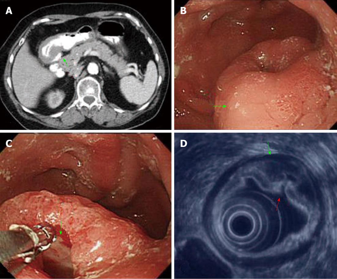
Figure 1 Image findings of gastric Peutz-Jeghers type stomach polyp.
A: Abdominal computed tomography scans of the patient revealing a thickened gastric wall over the antrum of the stomach (arrow); B: Gastroendoscopy revealing a bulging lesion (4 cm × 3 cm) of the antrum (arrow); C: The bulging lesion displayed a central hole (arrow); D: Endoscopic ultrasonography demonstrating that the appearance of a lesion was due to a thickened subepithelial layer (second layer lesion), submucosal layer thickened (third layer, green arrow), and corresponding hole (red arrow) over the antrum of the stomach.
- Citation: Jin JS, Yu JK, Tsao TY, Lin LF. Solitary gastric Peutz-Jeghers type stomach polyp mimicking a malignant gastric tumor. World J Gastroenterol 2012; 18(15): 1845-1848
- URL: https://www.wjgnet.com/1007-9327/full/v18/i15/1845.htm
- DOI: https://dx.doi.org/10.3748/wjg.v18.i15.1845


